
Oral Surgery
Surgical Tooth Extraction
in Chandigarh — Gentle Wisdom Tooth Removal by Dr. Sandhu
Impacted wisdom teeth, fractured roots, ankylosed teeth, root canal treated teeth — when a routine extraction is not sufficient, surgical technique is used to remove the tooth safely, preserve the surrounding bone, and set up the site for what comes next.
Duration
30–45 min
Pain Level
Minimal
Cost depends on the complexity of the case — tooth position, degree of impaction, whether bone removal or sectioning is required. All costs are agreed before treatment begins. No hidden charges..
- ★★★★★ 5-star rated on Google
- Verified reviews on Google
- Read Reviews →
— Surgical vs. Routine Extraction
When does an extraction become surgical?
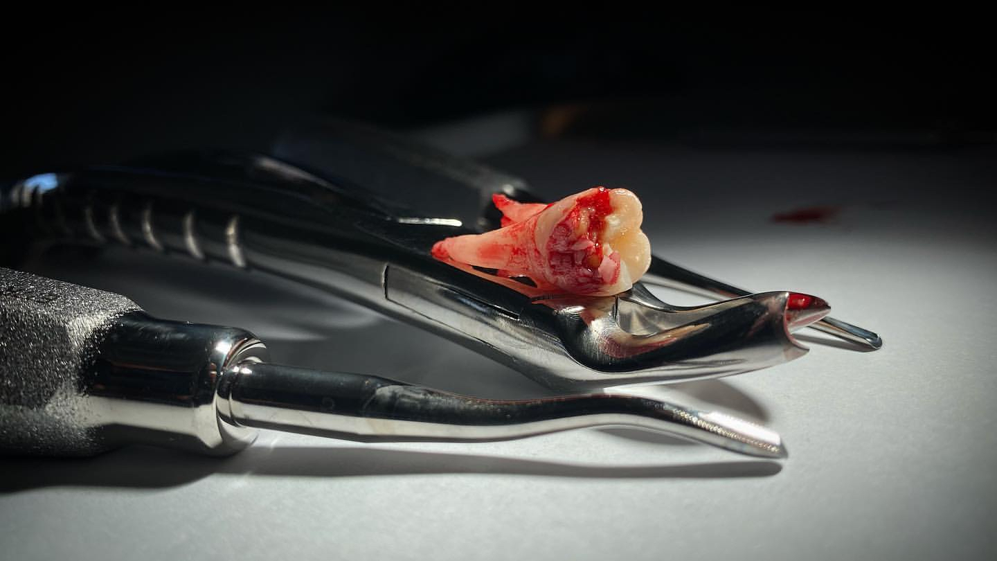
A routine extraction removes a tooth that is fully erupted and can be loosened from its socket with elevators and delivered with forceps. A surgical extraction is required when that pathway is blocked — when the tooth cannot be accessed, mobilised, or delivered without first raising the overlying gum and removing some of the surrounding bone.
The distinction matters because surgical extractions require a different level of planning, technique, and aftercare. At Tricity Smiles, every case is assessed with a periapical radiograph — and in the case of wisdom teeth, a detailed assessment of angulation, root morphology, and proximity to the inferior alveolar nerve — before a surgical approach is recommended.
If a tooth can be removed without surgery, it will be. The surgical approach is not a default — it is the correct choice when the anatomy requires it.
Indications
When surgical extraction is indicated
Four clinical situations that consistently require a surgical approach — each with specific challenges and surgical considerations.
3rd Molars
Impacted Wisdom Teeth
The degree of impaction determines the complexity: a partially erupted mesio-angular impaction requires less bone removal than a fully horizontal or vertical deep impaction. Proximity to the inferior alveolar nerve in the lower jaw is assessed on the pre-operative radiograph — cases where the roots are close to the nerve are managed with particular care to avoid post-operative numbness.
- Mesio-angular, disto-angular, horizontal, and vertical impactions
- Partially erupted — causing recurrent pericoronitis or decay
- Fully impacted — causing pressure on adjacent molar or cyst formation
- Lower wisdom teeth — proximity to inferior alveolar nerve assessed
Fractured Teeth
Broken or Fractured Teeth
Retained root fragments left in the socket are a source of chronic infection and a barrier to implant placement. At Tricity Smiles, retained roots are always addressed — not left in situ as a convenience. The fragment is identified on periapical radiograph, surgically exposed, and removed cleanly.
- Crown fracture during routine extraction — root retained in socket
- Subgingival fracture — no accessible purchase point above bone
- Curved or dilacerated roots that cannot be elevated without sectioning
- Retained root fragments from previous failed extractions
RCT Teeth
Root Canal Treated Teeth
Teeth with posts and cores are particularly challenging. The post bonds to the root, and forceps application to the crown transmits force differently than on an intact tooth. Surgical access — flap elevation and controlled bone removal — allows the root to be delivered with minimal lateral force and reduces the risk of leaving fragments in a site that may be destined for an implant.
- Failed root canal — periapical pathology persisting despite treatment
- Post and core present — elevated fracture risk during delivery
- Multiple rooted teeth with divergent or fused roots
- Pre-implant planning — surgical approach preserves the socket
Ankylosed Teeth
Ankylosed & Hypercementosed Teeth
Hypercementosis — excessive cementum deposition on the root surface creating a bulbous root shape — presents a similar challenge. The root cannot be delivered through the socket aperture without first surgically widening it or sectioning the root.
- Ankylosed deciduous teeth blocking permanent tooth eruption
- Ankylosed permanent teeth — confirmed by absence of PDL on radiograph
- Hypercementosed roots — bulbous root exceeding socket width
- Over-retained roots with significant bone ingrowth
The same thorough anaesthesia protocol — for every surgical case.
Topical gel first, pre-warmed articaine or lignocaine, the smallest appropriate needle, vibration technique, electronic delivery at a controlled rate, and aspiration to confirm placement. For lower wisdom teeth, a full inferior alveolar nerve block ensures profound numbness throughout. Nitrous oxide is available for anxious patients. Learn about nitrous sedation →
The Surgical Procedure
What happens during a surgical extraction
A methodical, planned procedure — each step preserving as much surrounding tissue and bone as possible.
01
Assessment & Anaesthesia
02
Flap Elevation
03
Bone Removal
04
Sectioning & Delivery
05
Socket Care
06
Flap Closure
— After the Tooth Is Out
Socket care, sterilisation, and what comes next
Surgical extraction produces a larger and more complex socket than a routine extraction — often with sutures, exposed bone edges, and a healing process that takes longer and requires more attentive aftercare.
Every instrument used during surgery is autoclaved to full sterilisation standards. Single-use items — needles, sutures, gauze, irrigation tubing — are discarded after every patient. The surgical field is maintained as clean as possible throughout the procedure to reduce the risk of post-operative infection — the most common complication of surgical extractions performed without proper sterile technique.
Sutures are typically removed or dissolve within 7–10 days. A follow-up appointment is scheduled to review healing, remove sutures where needed, and assess the socket before further treatment planning.
Planning an Implant?
Socket preservation at the time of surgical extraction
After a surgical extraction, the jawbone that supported the tooth root begins to resorb rapidly — and the resorption is often more significant than after a routine extraction because more bone has been disturbed during the procedure.
A socket preservation graft placed at the time of surgical extraction fills the void with graft material that maintains bone volume and architecture during healing. For premolars and molars where an implant may be placed subsequently, this step transforms a potentially complicated implant site into a predictable one.
For wisdom teeth, socket preservation is less commonly indicated — that space is rarely used for an implant. For any other surgical site where implant placement is being considered, the conversation about grafting should happen before the extraction, not after the bone has been lost.
Considering an implant to replace the extracted tooth?
— Why Tricity Smiles
Why surgical precision matters more than surgical speed
Pre-Operative Radiographic Assessment
Minimum Necessary Bone Removal
Saline Irrigation Throughout
US-Trained Surgical Judgement
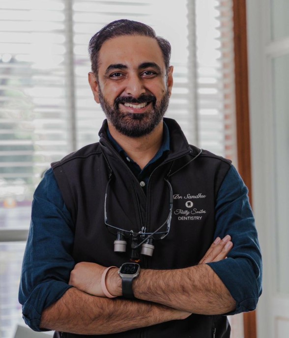
Dr. Sandhu
USC · UCSF · CMC Ludhiana
— Our Philosophy
The most important extraction is the one that preserves bone for what comes next.
A surgical extraction is rarely the end of the story. For most patients, the tooth being removed will eventually be replaced — with an implant, a bridge, or at minimum the space needs to be managed so adjacent teeth do not shift into it. How the extraction is performed determines how straightforward — or how complicated — the next chapter will be.
An extraction that removes bone unnecessarily — through overuse of the bur, aggressive socket curettage, or failure to graft — leaves a depleted site that requires additional surgery before an implant can be placed. An extraction that leaves root fragments in the socket leaves a source of chronic infection. An extraction performed without saline irrigation leaves bone cells damaged by heat, reducing the quality of the tissue that will eventually support a restoration.
Every extraction is a preparation for the next treatment. The site we leave behind determines what options the patient has going forward.
This is why surgical technique matters beyond simply getting the tooth out. Getting the tooth out is the minimum requirement. Getting it out in a way that preserves the maximum amount of bone, minimises soft tissue damage, maintains a clean surgical field, and leaves the patient with a socket that heals predictably — that is the standard we hold.
At Tricity Smiles, the planning conversation for a surgical extraction includes what happens after the surgery. If an implant is planned, we discuss socket preservation before the extraction appointment. If the extraction site will be left unrestored, we discuss what tooth movement may occur and when intervention should happen. The extraction is a single appointment — but the decisions made around it affect the patient’s oral health for years.
That is what we mean by treating the extraction as a preparation, not a conclusion.
— Pain Management After Surgery
Combination analgesics — no opioids.
First Line
Paracetamol 500mg–1g
Combined With
Ibuprofen (Combiflam) 400mg
— FAQ
Surgical extraction FAQs
Common questions about surgical tooth removal, wisdom teeth, and recovery in Chandigarh.
How much does wisdom tooth removal cost in Chandigarh?
Surgical extraction at Tricity Smiles starts from ₹8,000 per tooth. The final cost depends on complexity — tooth position, angulation, degree of impaction, and whether bone removal or sectioning is required. All costs are discussed and agreed before treatment begins.
Is surgical tooth extraction painful?
With thorough local anaesthesia, surgical extraction is not painful. We use the same detailed anaesthesia protocol as routine extractions — topical gel, pre-warmed solution, electronic delivery, vibration technique, and aspiration. For lower wisdom teeth, a full inferior alveolar nerve block ensures profound numbness. Nitrous oxide is available. You will feel pressure and movement — not pain.
What is an impacted wisdom tooth?
An impacted wisdom tooth is one that cannot erupt normally into the mouth — blocked by the adjacent molar, tilted within the bone, or fully embedded. It is removed surgically: the gum is reflected, bone removed to access the tooth, and the tooth sectioned and removed in pieces if needed. The degree of impaction determines how much bone removal and sectioning is required.
Why do RCT teeth sometimes need surgical extraction?
Root canal treated teeth are more brittle and at higher risk of fracturing during delivery. Teeth with posts and cores are particularly challenging. Surgical access — flap elevation and controlled bone removal — allows controlled delivery with minimal lateral force and reduces the risk of leaving root fragments in a site that may be planned for an implant.
What is an ankylosed tooth?
An ankylosed tooth has fused directly to the surrounding jawbone — there is no periodontal ligament separating them. Forceps application to an ankylosed tooth transmits force directly to the jaw without movement in the socket. Surgical extraction with bone removal is the only safe approach — confirmed on radiograph by the absence of a PDL space.
Should I get a bone graft when a wisdom tooth is removed?
For wisdom teeth, socket preservation is less commonly indicated as that space is rarely used for an implant. For surgical extractions of premolars or molars where an implant may be placed later, socket preservation at the time of extraction is strongly recommended — it maintains the bone volume needed and avoids more complex grafting later.
For Patients — Share This Link
After your surgical extraction
- Bite on gauze for 45–60 minutes — change if saturated, keep biting
- Apply ice pack to the cheek — 20 minutes on, 20 minutes off for 4–6 hours
- Take paracetamol and Combiflam alternately as prescribed — start before anaesthesia wears off
- Rest — keep head elevated, avoid physical exertion
- Do not rinse, spit, or use a straw for 24 hours
- Do not smoke — for at least 72 hours, ideally longer
- No hot food or drinks — eat soft, cool food on the opposite side
- Warm salt water rinses from Day 2 — 3–4 times daily, gently
- Soft diet for 5–7 days — avoid chewing on the surgical side
- Swelling peaks at 48–72 hours and then gradually reduces — this is normal
- Sutures dissolve or are removed at your follow-up appointment (7–10 days)
- Some oozing for 24–48 hours is normal — frank bleeding beyond this: call us
Severe pain from Day 3–4, or bleeding that won’t stop?
Related reading: Wisdom tooth removal explained
Removed carefully. Healed well. Ready for what’s next.
Book your assessment with Dr. Sandhu. Every surgical extraction at Tricity Smiles is planned to give the site the best possible foundation — whether for an implant, a bridge, or simply clean, predictable healing.
5★
Google Rating
5-star
rated on Google
20+
Years Experience